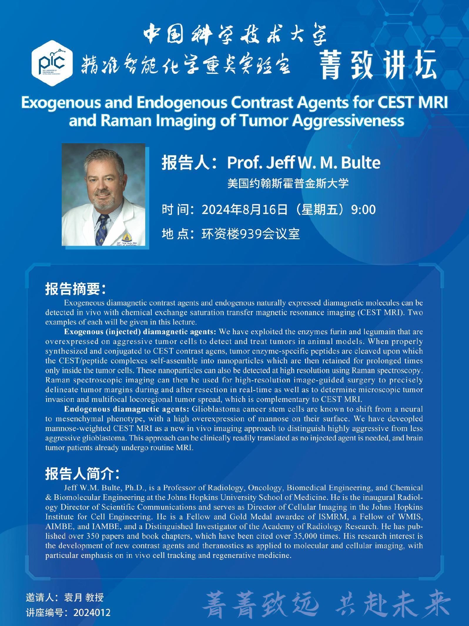
报告题目 | Exogenous and Endogenous Contrast Agents for CEST MRI and Raman Imaging of Tumor Aggressiveness |
报告人 | Prof. Jeff W. M. Bulte |
报告人单位 | 美国约翰斯霍普金斯大学 |
报告时间 | 2024年8月16日(星期五)9:00 |
报告地点 | 环资楼939会议室 |
主办单位 | 精准智能化学重点实验室 |
报告摘要 | Exogeneous diamagnetic contrast agents and endogenous naturally expressed diamagnetic molecules can be detected in vivo with chemical exchange saturation transfer magnetic resonance imaging (CEST MRI). Two examples of each will be given in this lecture. Exogenous (injected) diamagnetic agents: We have exploited the enzymes furin and legumain that are overexpressed on aggressive tumor cells to detect and treat tumors in animal models. When properly synthesized and conjugated to CEST contrast agens, tumor enzyme-specific peptides are cleaved upon which the CEST/peptide complexes self-assemble into nanoparticles which are then retained for prolonged times only inside the tumor cells. These nanoparticles can also be detected at high resolution using Raman spectroscopy. Raman spectroscopic imaging can then be used for high-resolution image-guided surgery to precisely delineate tumor margins during and after resection in real-time as well as to determine microscopic tumor invasion and multifocal locoregional tumor spread, which is complementary to CEST MRI. Endogenous diamagnetic agents: Glioblastoma cancer stem cells are known to shift from a neural to mesenchymal phenotype, with a high overexpression of mannose on their surface. We have deveopled mannose-weighted CEST MRI as a new in vivo imaging approach to distinguish highly aggressive from less aggressive glioblastoma. This approach can be clinically readily translated as no injected agent is needed, and brain tumor patients already undergo routine MRI. |
报告人简介 | Jeff W.M. Bulte, Ph.D., is a Professor of Radiology, Oncology, Biomedical Engineering, and Chemical & Biomolecular Engineering at the Johns Hopkins University School of Medicine. He is the inaugural Radiology Director of Scientific Communications and serves as Director of Cellular Imaging in the Johns Hopkins Institute for Cell Engineering. He is a Fellow and Gold Medal awardee of ISMRM, a Fellow of WMIS, AIMBE, and IAMBE, and a Distinguished Investigator of the Academy of Radiology Research. He has published over 350 papers and book chapters, which have been cited over 35,000 times. His research interest is the development of new contrast agents and theranostics as applied to molecular and cellular imaging, with particular emphasis on in vivo cell tracking and regenerative medicine. |

